

Parvoviral enteritis remains a major challenge in veterinary medicine today. Highly contagious, it primarily affects young unvaccinated animals and requires rapid and rigorous management.
In dogs, the causative agent is Canine Parvovirus (CPV), while in cats it is the Feline Panleukopenia Virus (FPV).
The main clinical signs are similar in both species: vomiting, diarrhea, dehydration, and leukopenia, all of which require immediate action.
Transmission occurs mainly via the faecal-oral route, but also via fomites (the environment, equipment and staff).
| Practical Overview | CPV (dog) | FPV (cat) |
| Main route of infection | Primarily fecal-oral transmission. | Primarily fecal-oral transmission. |
| Risk of silent infection | Early viral shedding, sometimes before clinical signs appear. | Early viral shedding, sometimes before clinical signs appear. |
| Critical role of the environment | Prolonged shedding: during illness and for 10–14 days after recovery (or longer). | Shedding is often shorter (1–2 days) but may last several weeks in some cats. |
| Viral load and contamination sources | Frequent indirect transmission through environment, equipment, and personnel. | Frequent indirect transmission through environment, equipment, and personnel. |
| Impact on the immune system | Frequent lymphopenia/neutropenia, which promotes bacterial translocation, sepsis and systemic complications. | Panleucopenia, which is often severe and sometimes profound, is associated with a more guarded prognosis. |
| Species-specific features | No major vertical transmission described. | In utero transmission possible, leading to fetal or neurological disorders in kittens. |
| Variable clinical expression | Highly resistant virus with prolonged environmental survival | Highly resistant virus with prolonged environmental survival |
Although closely related, CPV and FPV have both similarities and differences.
| CPV (dog) | FPV (cat) | |
| Early and marked vomiting | Upper gastrointestinal involvement | Frequent vomiting, often bilious |
| Often hemorrhagic diarrhea | Diarrhea / Intestinal involvement | Variable diarrhea, often non-hemorrhagic |
| Rapid progression within 24–48 hours | Disease progression | Acute course (5–7 days), hyperacute in kittens |
| Myocarditis possible in very young puppies | Age-related atypical forms | Neurological disease possible in kittens infected in utero |
| Lethargy, fever, dehydration | General condition | Profound lethargy, fever followed by possible hypothermia |
| Frequent leukopenia | Impact on immune system | Massive destruction of hematopoietic cells |
| Risk of septic shock | Severity and systemic complications | Risk of septic shock and complications (DIC, hypoglycemia) |
A key point: CPV most likely evolved from FPV, which explains their biological similarities.
When parvovirus infection is suspected, the challenge is not only to test, but to test appropriately and at the right time. Diagnosis should be considered as soon as the first compatible clinical signs appear, particularly vomiting and diarrhea in a young animal, lethargy, fever, leukopenia, or panleukopenia. Testing at the initial consultation allows rapid implementation of isolation measures and prompt initiation of treatment and supportive care.
|
|
In recently vaccinated animals, a positive result should be interpreted in light of the clinical context, as transient shedding of the modified live vaccine virus may occur.
Astéria® Parvovirus Feces and Astéria® Parvovirus Blood tests enable detection of parvovirus from either fecal or blood samples. This dual testing option offers a major clinical advantage: it allows diagnosis to be adapted to the stage of disease and the patient’s profile |
|

| Admission (Immediate Triage)
|
| Diagnosis: A Key Tool
|
| Patient Isolation
|
| Team Protection
|
| Care and Procedures
|
| Cleaning and Disinfection
|
| Management of Equipment and Traffic Flow
|
| After Hospitalization (Often Underestimated)
|
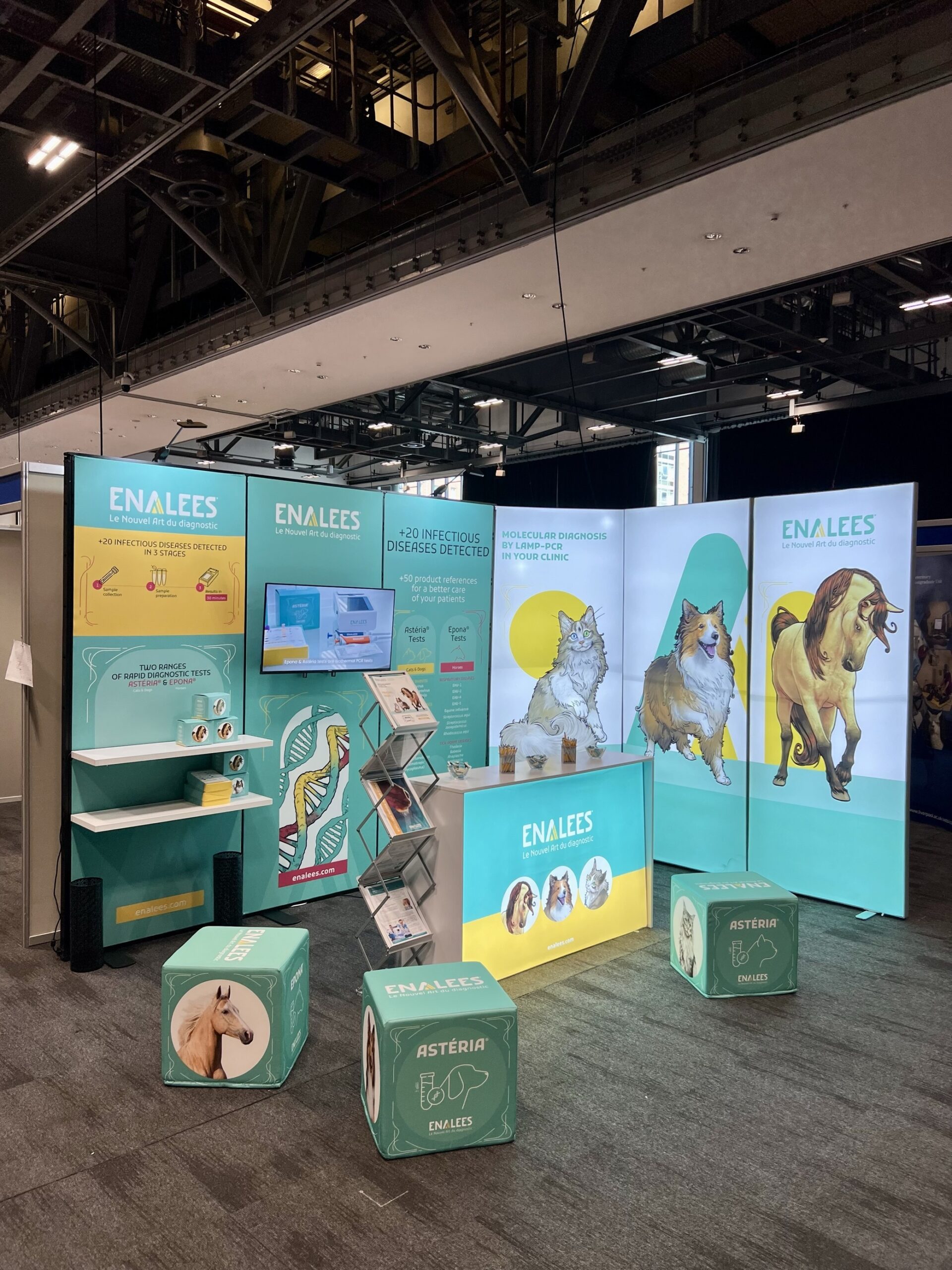
Throughout the year, Enalees takes part in the main conferences in the field of diagnostics, so that we can meet you, talk to you and present our results through posters and lectures!Here is a list of...
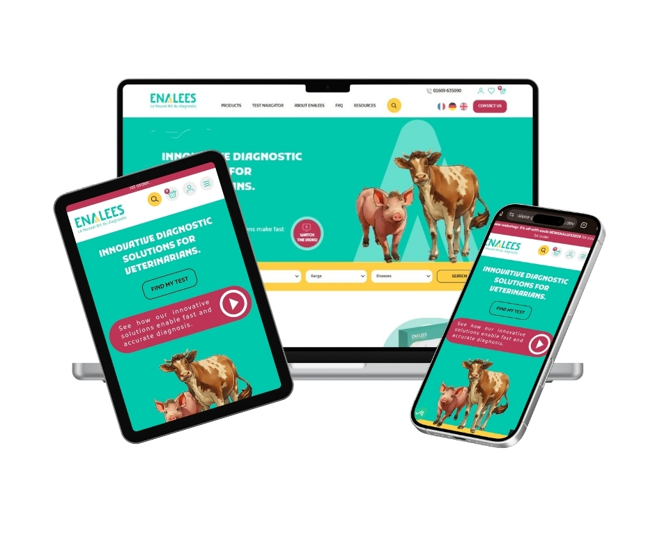
A central platform designed to organise clinic operations, make test selection easier and help teams focus their added value on clinical practice....
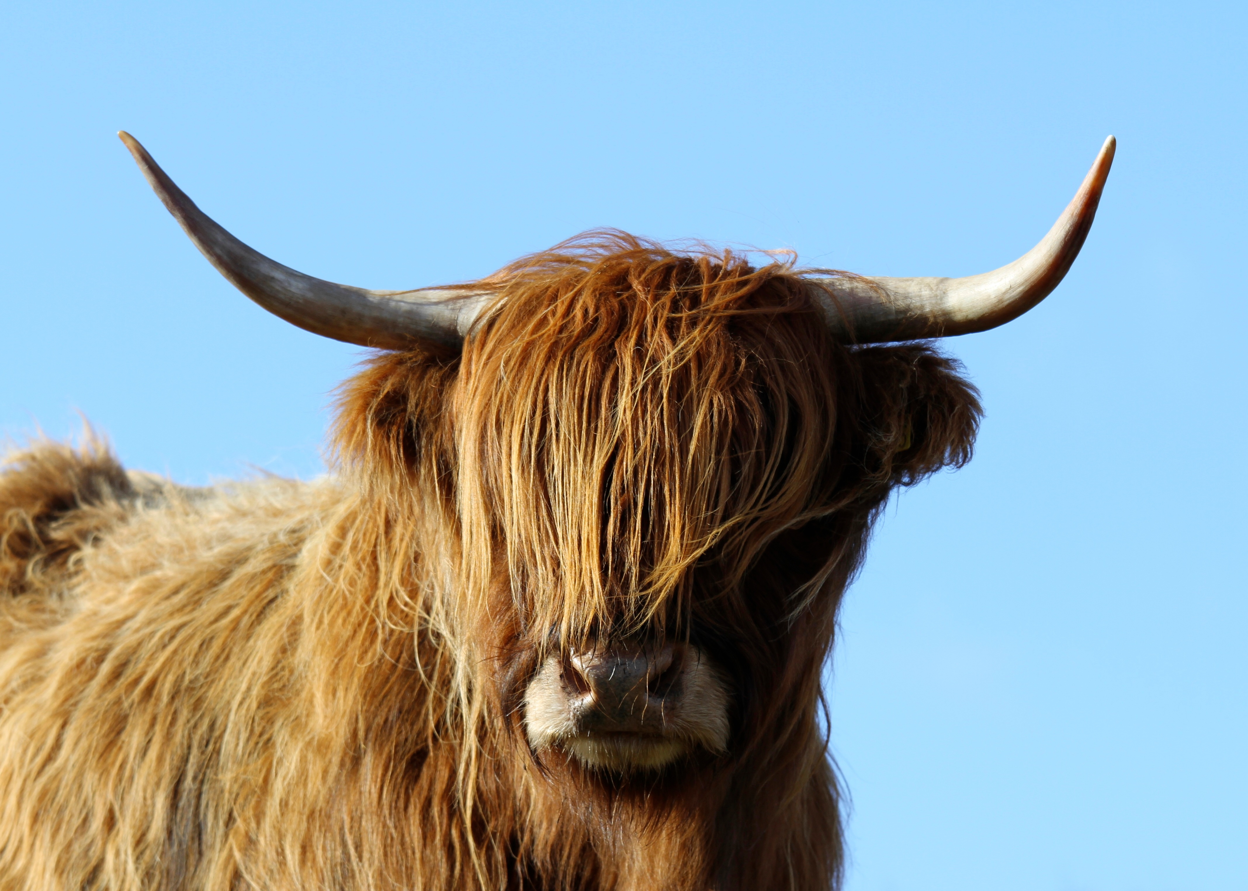
Bovine respiratory syncytial virus (BRSV) is a major cause of winter respiratory disease in cattle. Discover rapid diagnostic strategies....

Enalees Showcases Innovative LAMP Technology at AAEP Annual ConventionDecember 7th-11th, 2024 • Orlando (Florida) • Booth 1990We’re thrilled to announce our first participation at the ...
Enalees launches Astéria® Feline Coronavirus, a test designed to support veterinarians in the diagnosis of feline coronavirus in cats....